Maintaining regular check-ups, including cardiac imaging tests, is crucial to prioritising cardiovascular health. Cardiac imaging can come in various forms; with each using a different technique and offering different purposes.
Cardiac Imaging
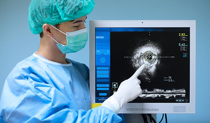
What Is Cardiac Imaging
Cardiac imaging is a test that uses specialised imaging techniques to visualise the heart, its surrounding tissue, and blood flow. The images produced allow the cardiologist to diagnose and monitor conditions affecting the heart, such as:
- Coronary artery disease
- Heart failure
- Congenital heart defects
- Heart valve problems
- Heart rhythm problems
When Do You Need a Cardiac Imaging Test?
A cardiac imaging test is recommended for individuals who have:
- Symptoms of Heart Disease – Imaging tests can help identify the underlying cause of symptoms such as chest pain, shortness of breath, fatigue, or palpitations. They can reveal structural abnormalities, blood flow issues, or other conditions that might be contributing to these symptoms.
- Risk Factors for Heart Disease – This test can detect high blood pressure, high cholesterol, and plaque buildup in the arteries. The earlier these risk factors are detected, the earlier treatments can be initiated to prevent further progression.
- Abnormal Test Results – If blood tests or electrocardiograms (ECGs) show concerning results, heart imaging tests can provide a clearer and more detailed picture and help the cardiologist determine the right treatment plan.
- Known Heart Conditions – Cardiac imaging tests are crucial for monitoring existing, known heart conditions. They help track the progression of the disease, evaluate the effectiveness of ongoing treatments and inform the cardiologist if adjustments to the treatment plan are necessary.
- A Scheduled Heart or Major Surgery – Cardiac imaging is often taken as part of the pre-surgical evaluation for heart or other major surgeries. It is used to evaluate the patient’s heart condition to plan the procedure and ensure the patient is cleared to undergo the procedure.
- Had a Heart Attack – After a heart attack, cardiac imaging is crucial to check for any damage to the heart muscle. It helps the cardiologist determine the best treatment for the patient to prevent further complications.
- Unexplained Syncope (Fainting) – These tests can help identify the presence of any heart-related causes behind fainting episodes.
Types of Non-Invasive Cardiac Imaging
Common types of non-invasive cardiac imaging include:
Echocardiogram
– Sometimes referred to as an echo, an echocardiogram uses ultrasound waves to create images of the heart. This test assesses the heart’s size, shape, valve function and blood flow to determine how effectively the patient’s heart works. It is also used to check for issues like heart valve disease, clots in the heart, and cancerous growths.Cardiac MRI
– Cardiac magnetic resonance imaging (MRI) is a painless, non-invasive imaging test that uses magnetic fields and radio waves to create detailed images of the heart and surrounding tissues. It offers comprehensive insights to help assess the heart’s function and identify scarring and other abnormalities. Cardiac MRI can help the cardiologist diagnose heart issues such as myocarditis, cardiac tumours, and pericardial diseases.Nuclear Stress Test
– A nuclear stress test evaluates blood flow to the heart muscle using imaging techniques. It involves injecting a small amount of radioactive material and monitoring blood flow during exercise or medication-induced stress. The test helps identify potential heart conditions, such as coronary artery disease.CT Coronary Angiography (CTA)
– CT coronary angiography is a non-invasive imaging test that uses computed tomography to visualise the coronary arteries. It helps identify blockages or abnormalities by injecting a contrast dye and capturing detailed images of the heart's blood vessels. This is used to assess coronary artery disease and guide treatment decisions.Cardiac PET Scan
– A cardiac Positron Emission Tomography (PET) scan uses radioactive tracers to look at how the heart and the surrounding tissues work in the body. When a patient has a heart attack, doctors may use a PET scan to determine which tissue in the patient’s heart is healthy and which parts are damaged.Multigated Acquisition (MUGA) Scan
– MUGA scans measure the heart's pumping function using a radioactive tracer. The radioactive tracer will be injected into the patient’s bloodstream and a special camera will create moving images of the beating heart. They take pictures at specific times during every heartbeat and produce a 2D image to see the dynamic properties of the heart and assess its structure.
How Long Does a Cardiac Imaging Test Take?
This depends on the type of test. Generally speaking, excluding preparation and waiting time, the actual imaging itself will take around:
- Echocardiography: 30-60 minutes
- Cardiac MRI: 45-90 minutes
- Cardiac CT: 10-30 minutes
- Nuclear Cardiology: 30 to 60 minutes
- Cardiac Catheterisation: 30-60 minutes
Risks Associated with Cardiac Imaging Tests
While cardiac imaging tests are considered safe and highly valuable for diagnosing heart conditions, and the benefits generally outweigh the risks, it may be helpful to take note of certain risks involved (as with any medical procedure):
- Allergic Reactions – Imaging tests such as coronary angiograms and CT scans involve the use of contrast dyes that can cause allergic reactions in rare cases.
- Radiation Exposure – Certain tests involve radiation exposure, which may be harmful in the long run depending on the dose and frequency (cumulative exposure) involved over time.
- Anxiety – The enclosed space of MRI scanners can cause discomfort or anxiety in people with claustrophobia.
FAQs
Cardiac imaging tests are generally safe and painless. Your cardiologist will take precaution to reduce any risks involved and keep you as comfortable and informed as possible.
This depends on the type of test performed and what the doctor wants to measure or assess. In general, the various cardiac imaging tests of today have high accuracy levels that can safely guide treatment and diagnosis.
Abnormal cardiac imaging results do not always mean a serious heart condition, but they do require a follow-up with your doctor. A cardiologist will be able to interpret the results in conjunction with your symptoms, medical history and other diagnostic tests; and determine the right course of action.
Our Insurance Partners
To keep our services accessible and convenient to our patients, we accept most major insurances
and can assist with the claims process.
We encourage you to call our clinic so we can review your coverage and assist you accordingly.


